Chylothorax in Dogs
Introduction
Chylothorax in dogs is an uncommon medical condition characterized by a buildup of chyle in the thoracic cavity, the hollow space where the lungs and heart are located. Due to this buildup, the lungs could have difficulty inflating when the dog breathes. Additionally, when not treated promptly, can lead to inflammation of the lungs and heart, as well as metabolic disorders and impaired immune system.
Pathogenesis of Chylothorax in Dogs
When food is digested, fats are further broken down into small molecules called chylomicrons. These fatty molecules are taken up by the dog’s lymphatic system. The chylomicrons mixed with lymphatic fluid makes up the chyle, a milky fluid that drains into the cisterna chyli, a structure in the abdomen, near the kidneys, which serves as a reservoir for this fluid.
From the cisterna chyli, the chyle is funneled into the thoracic duct so that it can be channeled back to the venous circulation. In dogs affected with chylothorax, chyle leaks from the thoracic duct into the chest cavity instead of being circulated into the bloodstream. Thus, aside from fluid buildup in the chest cavity, this disorder can also lead to a compromised immune system due to the substantial loss of lymphatic fluid in circulation.
Symptoms
Since the lungs are mainly affected by the fluid buildup, the signs and symptoms of chylothorax in dogs are often similar to other respiratory diseases characterized by an impediment to lung expansion like pneumothorax. Symptoms include:
- Breathing difficulty
- Non-productive cough
- Lethargy
- Rapid and shallow breathing
- Loss of appetite
- Weight loss
Over time, the dog may suffer from inflammation of the lungs and pericardium, compromised immune system, as well as irreversible damage to the lungs or heart, if left untreated.
Causes of Chylothorax in Dogs
Trauma or other underlying diseases of the circulatory system or thoracic organs like cancer, heart disease, and blood clots are some of the causes of this condition. These diseases could potentially impede the flow of chyle from the thoracic duct to the circulatory system. However, the majority of cases are idiopathic, which means the cause of the leak is unknown.
Diagnosis
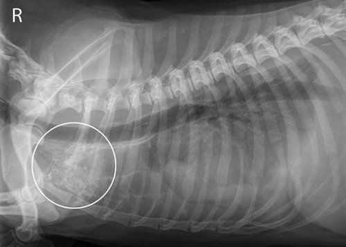
The veterinarian will first perform a physical exam of the pet. The thoracic area will be auscultated using a stethoscope to check for any abnormal sounds which are indicative of fluid buildup. When the veterinarian suspects fluid buildup, a thorax radiograph is needed to confirm and visualize the fluid inside the chest.
Once the presence of fluid is confirmed, a thoracentesis is necessary to determine what type of fluid is present in the chest cavity. This examination requires sedation of the dog before a small needle is inserted between two thoracic ribs to aspirate some fluid. If the fluid obtained has a milk-like appearance, it is a strong indication of chylothorax. The fluid is sent to the laboratory along with the dog’s blood test for a conclusive diagnosis of the disease.
The chylothorax could be a secondary complication of other health conditions so the veterinarian may recommend other diagnostic tests like computed tomographic (CT) scan as well as cardiac and thoracic ultrasound. These tests are more sensitive than the initial radiograph so the veterinarian can see clearly if there is a problem with the other organs that could be causing chylothorax.
Treatment
Treatment for chylothorax in dogs can be medical or surgical. When there’s an underlying condition that causes the leak, that medical condition is treated and medical management is employed to relieve the symptoms of chylothorax.
Medical management is a non-invasive treatment option that aims to reduce the formation of chyle and drain the chest cavity of fluid to relieve respiratory symptoms. A tube could be inserted into the dog’s chest and it will need to be admitted to the hospital, or the veterinarian can just decide to do an intermittent thoracocentesis.
The veterinarian may also recommend a switch to a low-fat diet for the dog to reduce the fat content of the chyle, although this approach has never been proven to reduce the amount of chyle leaked to the chest cavity. The dog may also be given fat-soluble vitamins to provide nutritional support for the dog.
The veterinarian will then check the dog and may recommend surgical intervention for these possible findings:
- There was no significant reduction in the volume of fluid in the chest cavity.
- The dog suffers from hypoproteinemia and protein-calorie malnutrition.
- The loss of chyle in circulation exceeds 20 ml/kg/day.
In these instances, surgery may be the best treatment option. Two surgical procedures often considered are:
Thoracic Duct Ligation
This surgical approach is mostly employed when dealing with idiopathic chylothorax in dogs. This method closes off the thoracic duct to encourage new lymphatic connections to the abdominal vein. In recent years, this procedure is done in conjunction with pericardiectomy or the removal of the heart lining. This is done since most chylothorax sufferers also exhibit a thickening of the heart lining, which could impede new lymphatic connections to the veins when the thoracic duct is ligated. A study reports a 90% success rate of thoracic duct ligation when performed with pericardiectomy.
Ablation of the Cisterna Chyli (CCA)
This surgical technique involves the removal of the cisterna chyli, the structure that serves as a reservoir of the chyle before it is channeled to the thoracic duct. The removal of this structure will force the body to adapt and create a new pathway to channel lymphatic fluids into the circulatory system. This will relieve pressure on the thoracic duct, reducing the volume of fluid leaked in the chest cavity.
In a study conducted involving 8 dogs, it was discovered that CCA combined with TDL significantly improved the success rate of the surgery, with all but one dog having fully recovered and free from symptoms of chylothorax.
While surgical treatment could be physically taxing for the dog, new techniques like the video-assisted thoracoscopy offer some relief as it helps surgical procedure become minimally invasive compared to the traditional open-chest surgery.
Cost to Treat: $6,000 to $10,000 when surgery is necessary
Aftercare and Prognosis
Post-operatively, recovery is most likely in the intensive care unit where the dog will be oxygenated and given medication for the pain. The chest tube inserted during surgery is aspirated intermittently to drain fluid from the chest cavity. The pet is most likely discharged when there’s no more effusion from the chest cavity and the tube can be removed.
Once discharged, pet owners will have to follow through with giving medication as prescribed. The veterinarian may also recommend a change of diet. Scheduled follow-up check-up to the vet should be observed as complications from the surgery like hemorrhage, nerve damage, infection, and persistent fluid accumulation in the chest could arise.
The reported success rate for thoracic duct ligation varies from 40% to 60%. If TDL is performed in conjunction with pericardectomy, improved success rates are reported at 80%-90%. TDL performed with CCA has a reported 88% success rate.
Treatment for chylothorax can be a lengthy process. Pet owners must cooperate with the veterinarian to provide the best care and support for dogs that went through surgery. And although this condition is very devastating and strenuous for pets and their owners, advancement in surgical techniques along with the use of minimally invasive technology offers relief and improved surgical success rate in dogs.
Sources:
- https://www.vetmed.wisc.edu/dss/ChyloThoraxTrial/page1.html
- https://www.acvs.org/small-animal/chylothorax
- https://www.ncbi.nlm.nih.gov/pubmed/15188816
Related Content


Compare Plans and Prices from the Top Companies