Dog Eye Ulcers
Corneal Ulcers in Dogs
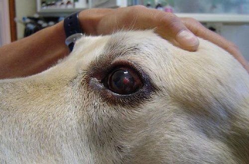
Dog eye ulcer, also known as corneal ulcer, happens when there is an erosion of the dog’s cornea. Majority of dogs that suffer from this condition experience superficial and non-infected corneal ulcers. Pets with this condition will develop clouded eyes due to fluid from its tears being absorbed into the deeper layer of the cornea known as the stroma.
Dog’s Cornea: An Overview
The cornea is a transparent and shiny film that covers the pupil and the iris. It makes up the entire front surface of the eyes and is made up of three layers, namely: the epithelium, stroma, and Descemet’s membrane. The epithelium makes up the outermost layer of the cornea. The stroma is the layer immediately below the epithelium. It’s the thickest layer and makes up approximately 90% of the corneas thickness because this serves as the supportive tissue. Lastly, the Descemet’s membrane is the deepest layer and it’s located directly above the aqueous humor, the fluid-filled space that separates the cornea from the lens.
Depending on which layer of the cornea is affected, dog eye ulcers can either be a minor problem or it can be a serious emergency that needs immediate intervention. In mild cases, only the epithelium could be affected in a condition known as a corneal abrasion or erosion.
Sometimes deeper erosion occurs, affecting the entire epithelium as well as parts of the stroma. In severe cases, erosion occurs not just in the epithelium and stroma, it could affect even Descemet’s membrane, resulting in a condition known as descemetocele. When the dog eye ulcer progresses to this stage, the Descemet’s membrane runs the risk of rupturing, causing the fluid in the aqueous humor to leak out and the eye to collapse. If left untreated, this could lead to irreparable damage.
Causes
The most common cause of corneal ulcers in dogs is trauma. Rough play with other dogs, running through a wooded area with lots of debris that could irritate the eyes, or constant rubbing of the eyes on the grass could lead to this condition. Also, when a cat scratches the dog’s eye or the eye comes in contact with a sharp object, this could cause a corneal laceration.
Chemicals can also cause corneal ulcers. The dog could be exposed to strong or irritating chemicals often found in some grooming products or from the environment. Genetics also plays a role in the development of this condition. Specifically, brachycephalic breeds like Pekingese, Shih Tzus, Pugs, Bulldogs, and Boxers are more susceptible to dog eye ulcers because of their shallow sockets providing less protection to their prominent eyes.
Furthermore, dog eye ulcer could be merely a symptom of other underlying conditions like:
Entropion. This condition occurs when the dog’s eyelids roll inward, causing the eyelashes and fur to constantly rub against the cornea.
Infection. Bacteria and viruses could release toxins that could irritate the eyes. Herpes virus and Salmonella are some of the viruses and bacteria that could cause ulceration of the cornea.
Dry Eye. Commonly referred to as keratoconjunctivitis sicca, this condition occurs when there’s an inflammation of the cornea along with other surrounding tissues due to inadequate tear production. Tears serve as a lubricant for the eyes and without an ample amount of it, the cornea could be irritated.
Endocrine Diseases. Dog eye ulcers could be one of the symptoms of medical conditions like Cushing’s disease or hyperadrenocorticism, diabetes mellitus, and hypothyroidism.
Epithelial Dystrophy. This hereditary condition is associated with breeds like Boxers, Shetland sheepdog, and Siberian Husky. Dogs that have this condition experience weakening of the cornea. Furthermore, lipid deposits could form at the superficial layer of the cornea, giving the eyes an opaque appearance.
Signs and Symptoms
The first thing pet owners will notice in dogs with eye ulcer is ocular pain and discomfort. Dogs will keep on pawing or rubbing their eyes. They may also keep on squinting or they may keep their eyes closed most of the time. Other signs include excessive tearing, redness, hypersensitivity to light, and the development of a thin film over the eyes.
Diagnosis
To diagnose this condition, the veterinarian will ask for the medical history of the pet. This is to establish probable causes for the dog’s eye ulcer. It’s also imperative that the vet knows the dog’s past or current medical conditions and the medications administered to it.
Furthermore, the veterinarian will perform an eye exam to inspect the cornea. But even with a thorough assessment, corneal abrasion may not be visible to the naked eye so the vet will have to use a special stain called fluorescein. Typically, water will just run off the surface of the cornea, but the fluorescent stain will adhere to the damaged surface of the cornea.
Other diagnostic tests like a complete blood count (CBC) and bacterial culture may also be needed. The CBC result could indicate or rule out infection. And if there is indeed an infection, the vet may request for bacterial or fungal culture and a subsequent antimicrobial susceptibility testing. This is done so that the vet can determine which antimicrobial regimen is most effective in treating the infection.
If an underlying medical condition is suspected, the vet will also pursue other laboratory tests to confirm or rule out his suspicion.
Treatment
Minor superficial and non-infected eye ulcers will usually resolve on its own after 3-10 days. But in most cases, treatment is necessary and two different treatment approaches or a combination of both are considered depending on the severity of the dog eye ulcer.
In cases where there’s a corneal abrasion or erosion, medications are prescribed to prevent infection and to relieve pain.
Antibiotics
A damaged cornea is always at risk for infection. If there’s already the presence of pus at the time of check-up with the vet, the cornea is already infected. That’s why the vet will prescribe an antibiotic eye drop or ointment to prevent or treat the infection.
Pain Reliever
The next part of the treatment is focused on pain relief. In many cases, 1% atropine sulfate eye drops or ointment is used for this purpose. This medication works by dilating the pupil since pupil spasm is the main source of pain for dogs with corneal ulcers. Atropine sulfate is known for its bitter taste and since the tear duct is connected to the mouth and nose, the dog may have a nasty reaction to it.
Corticosteroid
Normally, the cornea does not have blood vessels but when a descemetocele occurs, the body will try to promote healing by forming new blood vessels on the surface of the cornea. This process is called neovascularization. Although favorable for faster recovery, these blood vessels could impede vision. That’s why the vet may prescribe corticosteroid drops or ointment when the cornea has healed to reduce the size of these blood vessels.
Surgical
If the damage affects the deeper layers of the cornea, surgery might be the best recourse. The dead layers of the cornea will have to be removed in a procedure called grid keratectomy. In other cases, the veterinary ophthalmologist may consider replacing the damaged corneal tissue with healthy ones by performing a corneal graft. There are several types of corneal graft procedures but the commonly used are:
Conjunctival pedicle graft
The conjunctiva, the pinkish tissue that lines the inside of the eyelid, is rich in blood vessels. In this procedure, some conjunctival tissues are harvested and sutured into the defective corneal tissue. The conjunctival tissue will not just act as supportive tissue, but it will also provide the much-needed blood supply so the cornea can heal faster. This surgical procedure is suitable for deep yet small dog eye ulcers.
Corneo-conjunctival transposition graft
In this procedure, healthy corneal tissues adjacent to the damaged ones are harvested. These tissues, along with attached conjunctival tissues, are sutured into the damaged tissue. This procedure is suitable for deep and large ulcers. The only downside is that this may not be suitable when there is severe corneal infection already.
Pet owners need to have an extensive discussion with the ophthalmologist if they’re considering a surgical procedure for their pet.
Aftercare
A week after therapy, the dog will have to undergo another fluorescein test so the veterinarian can assess the rate of healing. While going through the therapy, the dog has to be closely monitored. If the dog underwent surgery or is given eye drops or ointment, it will have to use an Elizabethan collar to prevent self-injury and facilitate the fast healing of the affected eye.
Pet owners should check any irregularities with the affected eye or if there are unusual discharges. Additionally, activity should be kept at a minimum to prevent trauma to the eyes and medications are administered as prescribed.
Simple corneal ulcers should heal in about 1-2 weeks. If the cornea isn’t showing any sign of progress within this time frame, there may be an underlying condition that wasn’t addressed during treatment. Pet owners need to schedule a consultation with the veterinarian right away to determine the inciting cause.
Sources:
- https://vetmed.illinois.edu/refractory-corneal-ulcer-management-dogs/
- https://www.vin.com/apputil/content/defaultadv1.aspx?pId=11242&catId=31933&id=3860707
- https://cidd.discoveryspace.ca/disorder/corneal-dystrophy.html
Related Content


Compare Plans and Prices from the Top Companies