Glaucoma in Dogs

Overview
Glaucoma defines a group of eye conditions that are characterized by an elevated intraocular pressure, which typically leads to optic nerve damage. It’s a painful condition and the most common cause of blindness in dogs.
What is Intraocular Pressure?
The shape and size of the eyeball is regulated by fluid known as aqueous humor. Essentially, the eye is a fluid-filled ball, and the pressure of this fluid is known as intraocular pressure (IOP).
Eye fluid is constantly produced and contains nutrients and oxygen necessary for the eye to function optimally. Excess fluid is released through the “drainage angle” to maintain optimal IOP.
If fluid does not drain effectively, the IOP rises. Subsequently, the eye often gets larger and may take on an irregular shape. If not addressed immediately before this happens, the increased pressure can damage the optic nerve, which typically leads to vision loss.
High pressure is what causes pain, which most likely resembles a headache or even a migraine.
Early Warning Signs and Symptoms
Early intervention and treatment are necessary for the best possible outcome. Therefore, it’s extremely important to recognize the initial signs and symptoms.
At the onset of glaucoma, your dog may experience one or more of the following:
- Red and/or cloudy eye(s)
- Bloodshot eye(s)
- Abnormally small or large pupil(s)
- Abnormal blinking
- Squinting or fluttering eyelids
- Loss of appetite
- Lack of interest in playing or socializing
- Rubbing eye with paw or on the floor (due to pain)
- Tearing or watery discharge
- Crust around eye(s)
- Bumping into walls and furniture
- Swollen eye(s)
- One eye larger than the other
- Intolerance to light
- Third eyelid elevation
Types of Glaucoma
Glaucoma is classified as either primary or secondary.
1. Primary Glaucoma
Primary glaucoma often occurs suddenly without warning. It’s believed to be genetic and caused by a physical or functional abnormality that prevents eye fluid from draining properly. For example, the drainage angle may be deformed or the opening may be too small.
Primary glaucoma typically starts in one eye and then moves to the other. Thus, always check for differences between the two eyes.
The age of onset is usually between four and ten years. However, it is possible at any age.
While primary glaucoma has been documented in mostly all dog breeds, some breeds are predisposed, which include:
- Cocker Spaniel
- Basset Hound
- Wire Fox Terrier
- Boston Terrier
- Chow Chow
- Shar-Pei
- Norwegian Elkhund
- Siberian Husky
- Samoyed
- Cairn Terrier
- Maltese
- Miniature Poodle
- Beagles
- Dalmatian
- Chihuahua
- Vizsla
In North America, the prevalence of primary glaucoma in these predisposed breeds can be as high as 5.5%.
2. Secondary Glaucoma
Secondary glaucoma occurs when IOP increases as a result of another eye related disease or damage to the eye. Such conditions include:
- Cataracts
- Tumors
- Lens dislocation
- Penetration of the eye
- Infection
- Inflammation (anterior uveitis)
- Scarring from injury
- Bleeding and blot clots
- Retinal detachment
As a result, diagnosis and treatment of these conditions is also time sensitive to prevent the occurrence of glaucoma. Further, dogs with these disorders should routinely have their IOP measured.
While secondary glaucoma is not considered hereditary, the underlying conditions may be, such as cataracts and lens dislocation.
The combined prevalence of primary and secondary glaucoma is approximately 2% in the general canine population.
It’s also worth noting that the prevalence of cataracts (a common cause of secondary glaucoma) is estimated to be as high as 3.5% within the general canine population.
However, for breeds with genetic predispositions, the prevalence has been calculated as high as 11%. Further, several of the same breeds predisposed to primary glaucoma are also more likely to develop cataracts, including cocker spaniel, terriers, and miniature poodle.
Inflammation caused by anterior uveitis is also very common in dogs.
Diagnosis
As previously mentioned, immediate medical treatment is necessary if your dog exhibits one or more of the symptoms listed above. Complete loss of vision is more likely to occur the longer you wait. In some cases, blindness can occur within hours.
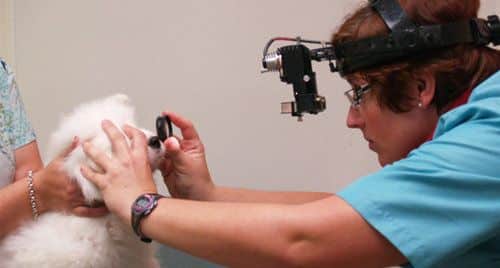
The veterinarian will perform an eye examination as well as measure the fluid pressure with an instrument known as a tonometer.
For a majority of dogs, a normal IOP is between 15 and 25 mmHg. Early stages of glaucoma typically produce IOP results between 20 an 30 mmHg. Moderate cases reach IOP levels between 30 mmHg and 40 mmHg. With advanced stages, IOP can rise between 40 and 50 mmHg.
If the pressure is raised above normal, and there are no other obvious explanations, glaucoma is diagnosed.
Promptly you will need to see a veterinary ophthalmologist who has all the necessary equipment to further evaluate the eye to determine the best course of action.
Specifically, the ophthalmologist uses special tools to examine the drainage angle and the optic nerve. X-rays and ultrasounds may also be required to rule out the presence of tumors, injuries, and abscesses.
Treatment Options
Treatment depends on the severity of damage, and each case is different.
In general, the key goals of treatment are to:
- Reduce pain
- Reduce intraocular pressure
- Increase drainage
- Decrease fluid production
If there’s a chance to save your dog’s vision, medical treatment will be administered to reduce IOP. The drug or combination of drugs your ophthalmologist chooses with depend on the level of pressure and condition of the optic nerve.
Some medications are given orally, while others get placed directly in the eye. The most commonly prescribed drugs include:
- Osmotic diuretics – reduce fluid production
- Carbonic anhydrase inhibitors – reduce fluid production
- Miotics – promote fluid release by constricting the pupil
- Adrenergic drugs – promote fluid release
- Prostaglandin analogs – promote fluid release
- Beta – blockers – reduce fluid production
With secondary glaucoma, the underlying disease or dysfunction must also be treated. This may involve medications to reduce inflammation (i.e., corticosteroids) or treat an infection (i.e., antimicrobials).
While medication can reduce pain and delay disease progression, it’s not an effective long-term solution. Thus, once IOP has been reduced, surgery is most often necessary.
Some surgical procedures aim to reduce fluid production. However, other more successful procedures involve the use of implants to promote better fluid drainage. Specifically, a small hollow tube is placed in the eye to prevent blockages.
In either case, success is not guaranteed and complications are possible. Thus, the eye will need to be carefully monitored on a regular basis. Unfortunately, repeat surgeries may be necessary.
Further, certain medications (i.e., cholinesterase) may be prescribed to slow the progression of disease in the unaffected eye. As previously mentioned, primary glaucoma almost always occurs in both eyes.
In the case of irreversible vision loss, which occurs in approximately 40% of cases, surgical removal of the eye is recommended. It’s truly the best way to alleviate pain and prevent further complications.
Cost of Treatment
As discussed above, primary glaucoma can develop suddenly without warning. Further, treatment is required immediately after diagnosis.
Below is a summary of the expenses you will most likely incur if your dog is diagnosed with glaucoma:
- Emergency veterinary hospital visit
- Ophthalmologist office visit
- Eye examinations (i.e., IOP measurement, optic nerve imaging, ultrasound, X-rays)
- Medications (immediate and ongoing)
- Surgery (possibly more than one)
- Follow-up office visits
In addition, if your dog’s vision is lost, you may need to make certain modifications in your home to ensure his or her safety. Needless to say, the cost of treatment is high. It can reach as much as $3,500 in a very short period of time.
Avg. Cost of Treatment: $2,000 to 3,000
Treatment of glaucoma in dogs may be medical or surgical. Treating serious eye problems like glaucoma can cost thousands of dollars. Pet health insurance helps pay for expensive veterinary care if your dog gets sick or injured.
Conclusions
- Glaucoma affects the eye and is one of the leading causes of blindness in dogs
- Identifying early warning signs is essential since the disease progresses quickly
- Prompt treatment is necessary and most often involves medications as well as surgery
- Cost of immediate and ongoing treatment is high
Related Content:
References
Gelatt, K. N. (2014). Essentials of veterinary ophthalmology. Ames, IA: Wiley Blackwell.
Gelatt, K. N. et al. (2005). Prevalence of primary breed-related cataracts in the dog in North America. Veterinary Ophthalmology, 8(2), 101-111. doi:10.1111/j.1463-5224.2005.00352.x
Gelatt, K. N. et al. (2004). Prevalence of the breed-related glaucomas in purebred dogs in North America. Veterinary Ophthalmology, 7(2), 97-111. doi:10.1111/j.1463-5224.2004.04006.x
Gelatt, K. N., at al. (2004). Secondary glaucomas in the dog in North America. Veterinary Ophthalmology, 7(4), 245-259. doi:10.1111/j.1463-5224.2004.04034.x
Johnsen, D. A., et al. (2006). Evaluation of risk factors for development of secondary glaucoma in dogs: 156 cases (1999–2004). Journal of the American Veterinary Medical Association, 229(8), 1270-1274. doi:10.2460/javma.229.8.1270
Mellersh, C. S. (2014). The genetics of eye disorders in the dog. Canine Genetics and Epidemiology,1(1), 3. doi:10.1186/2052-6687-1-3
Miller, P. E., et al. (2015). Clinical Signs and Diagnosis of the Canine Primary Glaucomas. The Veterinary Clinics of North America. Small Animal Practice, 45(6), 1183–vi. http://doi.org/10.1016/j.cvsm.2015.06.006
Researchers Advance New Glaucoma Treatments. (2015, February 3). Retrieved March 10, 2017, from https://cvm.ncsu.edu/researchers-advance-new-glaucoma-treatments/
Tinsley, David M., et al. (1993) Glaucoma: Past and Present Management Techniques. Iowa State University Veterinarian, 55(1).


